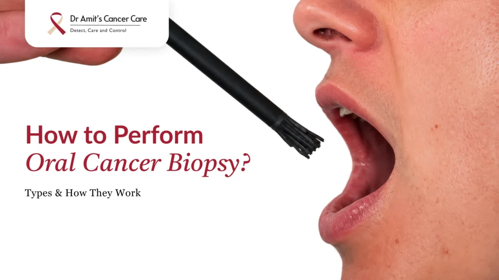

Oral cancer biopsy is one of the tests done to detect cancer. It is done when you find yourself suffering from a white patch or lump inside your mouth that just won’t heal. The procedure is carried out by excising some part of the tissue and examining the cells to check for oral cancer.
The mouth biopsy procedure is done by a doctor, and the cells, when examined, may be benign (non-cancerous) or malignant (cancerous), which helps diagnose oral cancer.
This guide walks you through how an oral cancer biopsy is performed, the different oral biopsy techniques used, what to expect during recovery, how to understand your oral biopsy results, and when to consult based upon your mouth cancer biopsy reports.
What Is an Oral Cancer Biopsy?
An oral cancer biopsy, also known as a mouth cancer biopsy, is a minor procedure where a small piece of tissue is removed from the mouth and sent to a lab for examination under a microscope.
This biopsy of oral tissue is only carried out when there is a lump or a painless swelling that is suspected of mouth cancer. It is one of the widely practised methods for confirming whether a suspicious white patch, red patch, ulcer, or lump is benign, precancerous, or cancerous.
While imaging and visual exams may only suspect cancer, only a proper biopsy of the mouth, examined by a pathologist, can give an exact diagnosis (Anjum et al., 2024).
If you’ve noticed a persistent sore or growth in your mouth, it is recommended to schedule an oral cancer screening before symptoms progress.
Who Needs an Oral Cancer Biopsy?
A mouth cancer biopsy may be needed when there is an ulcer or a discoloured patch that is non-healing in nature for more than 2 weeks.
Not every mouth ulcer needs a biopsy. Most of the oral ulcers heal on their own within 1–2 weeks.
An oral ulcer biopsy is generally recommended when:
- A sore or ulcer lasts more than 2–3 weeks without healing
- There’s a white or red patch that won’t rub off
- You notice an unexplained lump, swelling, or thickened area
- There’s unusual bleeding, numbness, or pain in the mouth with no obvious cause
- A lesion looks suspicious on visual or adjunctive screening (such as toluidine blue staining)
If any of these sound familiar, don’t wait. Early detection dramatically improves treatment outcomes, and so it is important to be aware of the warning signs of oral cancer.
If you or your loved one is suffering from any of these symptoms, early detection is recommended. Book a consultation for a detailed assessment from Dr. Amit Chakraborty.
A persistent oral ulcer, unexplained patch, lump, or other unusual mouth changes should be evaluated without delay. Dr. Amit Chakraborty and his team provide a detailed assessment of suspicious oral lesions and can guide you on whether a biopsy or further evaluation is required.
Oral Biopsy Techniques
There are many techniques of mouth biopsy, including incision, excision, punch, fine-needle and brush.
The best oral biopsy technique is recommended based upon the size, location, and appearance of the lesion.
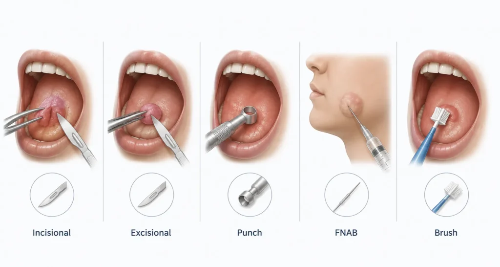
Following are the main mouth biopsy techniques:
1. Incisional Biopsy
Only a small portion of the lesion is removed, along with a bit of surrounding tissue. This is the preferred technique when a lesion is large, appears suspicious for cancer, or is in a sensitive location since it allows diagnosis without removing the entire area. (Golden and Hooley, 1994; Anjum et al., 2024).
2. Excisional Biopsy
The entire lesion is removed, usually along with a small margin of healthy tissue around it. This method is typically used for smaller lesions (generally under 1 cm).
3. Punch Biopsy
A specialised circular punch tool is used to remove a small cylindrical piece of tissue. It’s useful for hard-to-reach areas, though it’s used less often inside the mouth compared to the skin, since it may harm delicate oral tissue.
4. Fine Needle Aspiration Biopsy (FNAB)
A thin needle is used to draw out a small sample of cells, most often used for lumps in the salivary glands or swellings under the jaw, rather than surface lesions.
5. Brush Biopsy
A small brush is used to collect surface cells for an initial screen, which is useful as a preliminary check, though it’s not a substitute for a full tissue biopsy if cancer is strongly suspected.
Doctors often combine a visual exam with toluidine blue dye to pinpoint exactly where the biopsy should be taken from, which improves diagnostic accuracy (Anjum et al., 2024).
Dr. Amit Chakroborty personalises the choice of technique based on your specific lesion. You can learn more in his guide to diagnostic tests for oral cancer.
Oral Biopsy Procedure
The procedure of oral biopsy is quick and safe, where a doctor numbs the area with local anaesthesia, removes a small piece of abnormal mouth tissue, and sends it to a lab for testing. The whole process takes about 15 to 20 minutes, and you may feel pressure instead of sharp pain.
Here’s exactly what happens during the mouth biopsy procedure, from start to finish:
| Step | What Happens During the Biopsy? |
| Clinical Examination | The doctor examines the lesion, noting its size, location, colour and texture, while also reviewing your symptoms and medical history. |
| Local Anaesthesia | A numbing injection is given around the biopsy site to make the procedure as comfortable and pain-free as possible. |
| Stabilising the Tissue | The lesion and nearby tissue are gently held with special non-toothed forceps to avoid damaging the sample. |
| Taking the Tissue Sample | Depending on the biopsy type, either the entire lesion or a small representative portion is carefully removed using a scalpel, punch or laser. |
| Controlling Bleeding | Bleeding is managed using gentle pressure, stitches (sutures), or cautery to ensure the area is safe and secure. |
| Closing the Wound | Most biopsy sites are closed with a few dissolvable stitches to promote proper healing. |
| Preserving the Sample | The tissue is immediately placed in a preservative solution (usually 10% formalin) before being sent to the laboratory for analysis. |
| Laboratory Analysis | A pathologist examines the tissue under a microscope and prepares a detailed report to confirm the diagnosis and identify the exact condition. |
The entire in-clinic procedure usually takes just 15–30 minutes. If you’re due for one, Dr. Amit Chakraborty’s pre-biopsy preparation checklist explains exactly how to prepare for your appointment.
Oral Biopsy Recovery
Recovery from a mouth tissue biopsy generally takes one to two weeks. It is important to manage by avoiding hot and spicy food as well as spitting for 24 hours. With this, activities such as smoking or tobacco should also be avoided.
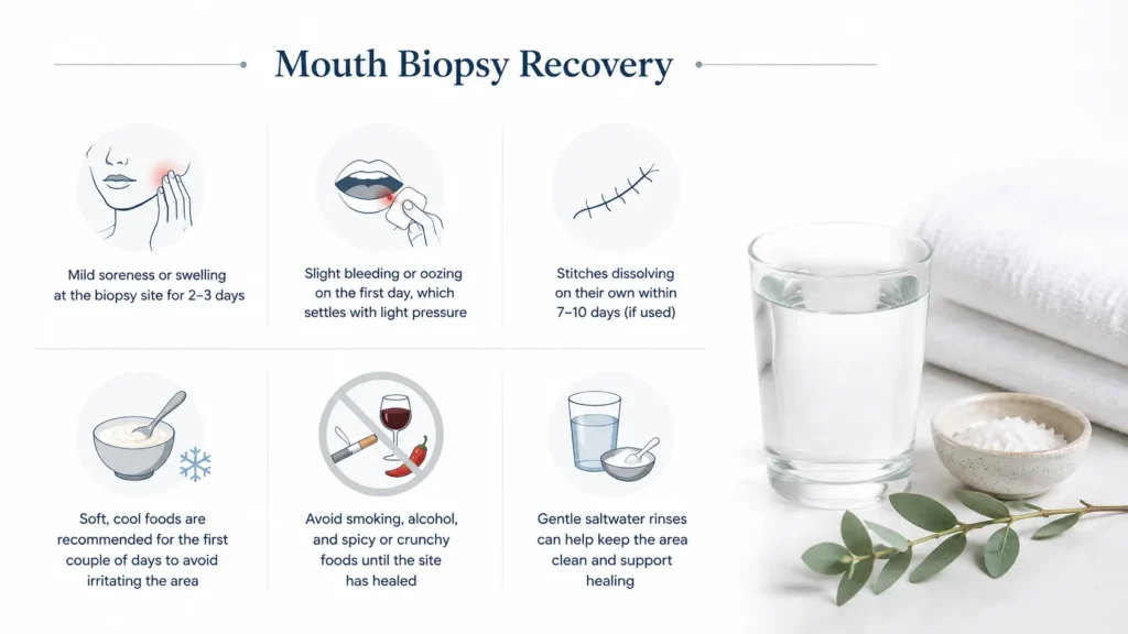
Mouth biopsy recovery is usually quick and manageable. Following are the recovery instructions to be taken care of:
- Mild soreness or swelling at the biopsy site for 2–3 days
- Slight bleeding or oozing on the first day, which settles with light pressure
- Stitches dissolving on their own within 7–10 days (if used)
- Soft, cool foods are recommended for the first couple of days to avoid irritating the area
- Avoid smoking, alcohol, and spicy or crunchy foods until the site has healed
- Gentle saltwater rinses can help keep the area clean and support healing
Most people are back to their normal routine within a day or two. If you notice heavy bleeding, worsening pain, fever, or pus, contact your doctor promptly. Dr. Amit Chakraborty’s team provides detailed post-biopsy care instructions to every patient to make recovery as smooth as possible. Lear more about the importance of oral cancer biopsy from Dr. Amit Chakraborty.
क्या Oral cancer केलिए biopsy जरुरी है? | Muh Ke Cancer Ka Test | Dr Amit Chakraborty, Mumbai
Understanding Your Oral Biopsy Results
An oral biopsy report details the microscopic analysis of a tissue sample taken from your mouth. It confirms if a lesion (such as an ulcer, lump, or white patch) is benign or malignant and identifies conditions like oral submucous fibrosis or leukoplakia.
Waiting for mouth biopsy results can be one of the hardest parts of the process. Your report of the oral biopsy will generally fall into one of these categories:
| Biopsy Result | What It Means |
| Benign | No cancerous or precancerous cells are found. The lesion is non-cancerous and usually does not require cancer treatment, though routine follow-up may be recommended. |
| Dysplasia (Precancerous Changes) | Abnormal cells are present that are not yet cancer, but they have the potential to develop into cancer over time. Regular monitoring or further treatment may be recommended to prevent progression. |
| Malignant (Cancerous) | Cancer cells are confirmed in the biopsy. Your doctor will discuss the next steps, which may include additional tests, cancer staging, and a personalised treatment plan. |
Next Steps
Whatever the outcome, it’s important to review your results with your doctor rather than trying to interpret them alone, since oral cancer biopsy reports use technical pathology terms that need clinical context.
Dr. Amit Chakraborty, a specialised head and neck oncosurgeon in Mumbai, offers a dedicated results consultation session to walk you through your report clearly and answer every question you have.
If you’ve been advised to get a biopsy or you’re concerned about a lesion that isn’t healing, book a consultation with Dr. Amit Chakraborty for an accurate, timely diagnosis.
Understanding your biopsy report can be difficult without the right medical context. Dr. Amit Chakraborty and his team can help you understand your results, explain what the findings mean, and guide you through the appropriate next steps.
References
- Anjum, R., Raj, R., Vijay, P., Kouser, A., Singh, P. and Jehan, A., 2024. Biopsy techniques: a review. Journal of Oral Medicine, Oral Surgery, Oral Pathology and Oral Radiology, 10(3), pp.181–184.
- Golden, D.P. and Hooley, J.R., 1994. Oral mucosal biopsy procedures. Excisional and incisional. Dental Clinics of North America, 38(2), pp.279–300.
- Yang, G., Wei, L., Thong, B.K.S., Fu, Y., Cheong, I.H., Kozlakidis, Z., Li, X., Wang, H. and Li, X., 2022. A systematic review of oral biopsies, sample types, and detection techniques applied in relation to oral cancer detection. BioTech, 11(1), p.5.


 May 22, 2026